Sinus lifting is a surgical procedure aimed at increasing the bone volume in the upper jaw to allow for the successful placement of implants. This method, which has recently emerged in dentistry but has already shown impressive results, improves the bone structure, making it possible to successfully insert titanium rods for subsequent crown attachment.
In the past, patients with insufficient bone volume in the upper jaw often could not receive implants and had to rely on bridge constructions or removable dentures. However, with the advent of sinus lifting, this situation has changed. Now, implants can be placed in almost all patients, despite initial limitations.
There are two types of sinus lifting: vertical and lateral. The latter, most common, is performed through a lateral approach to the maxillary sinuses, allowing for a significant increase in bone volume.
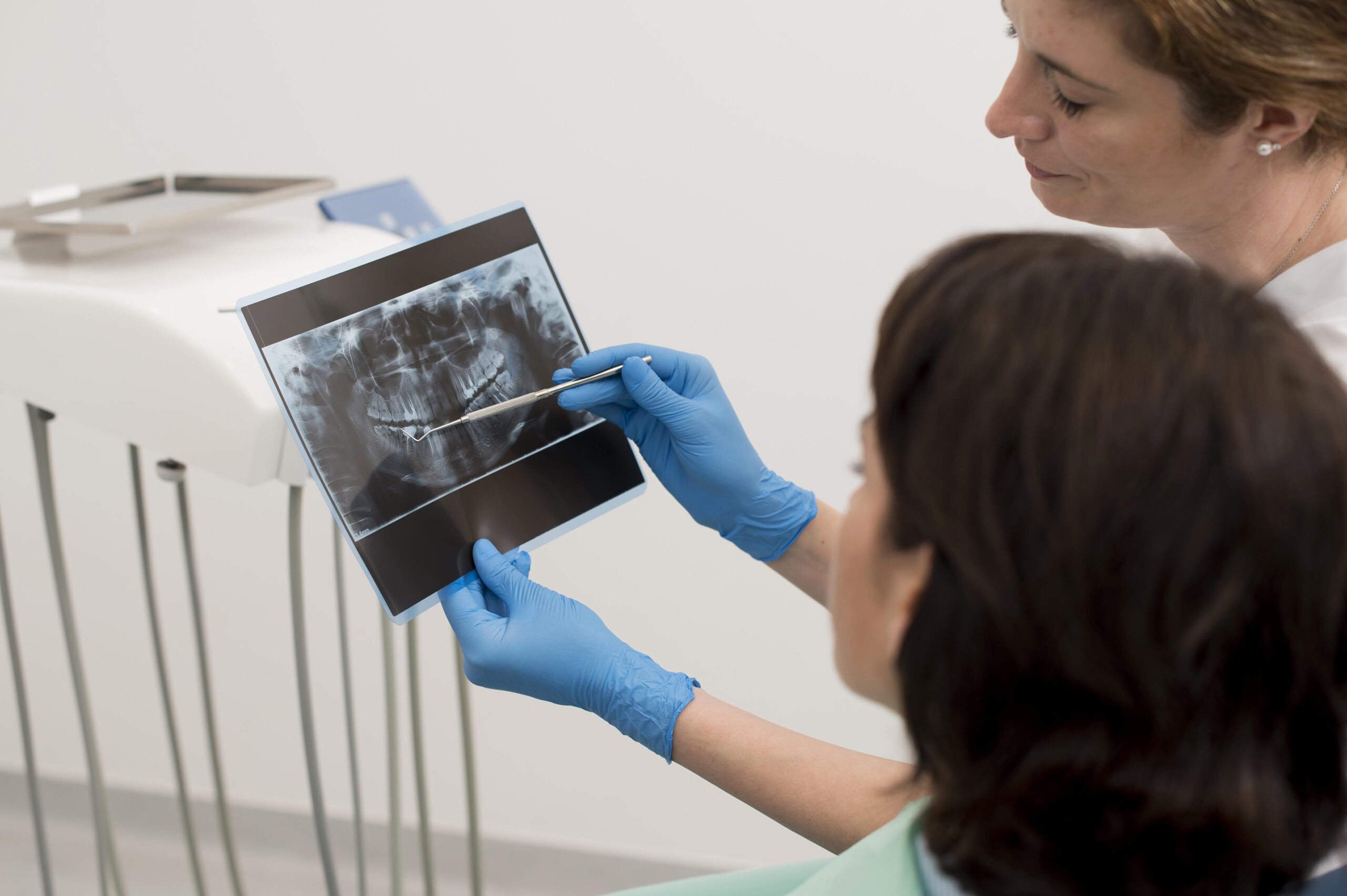
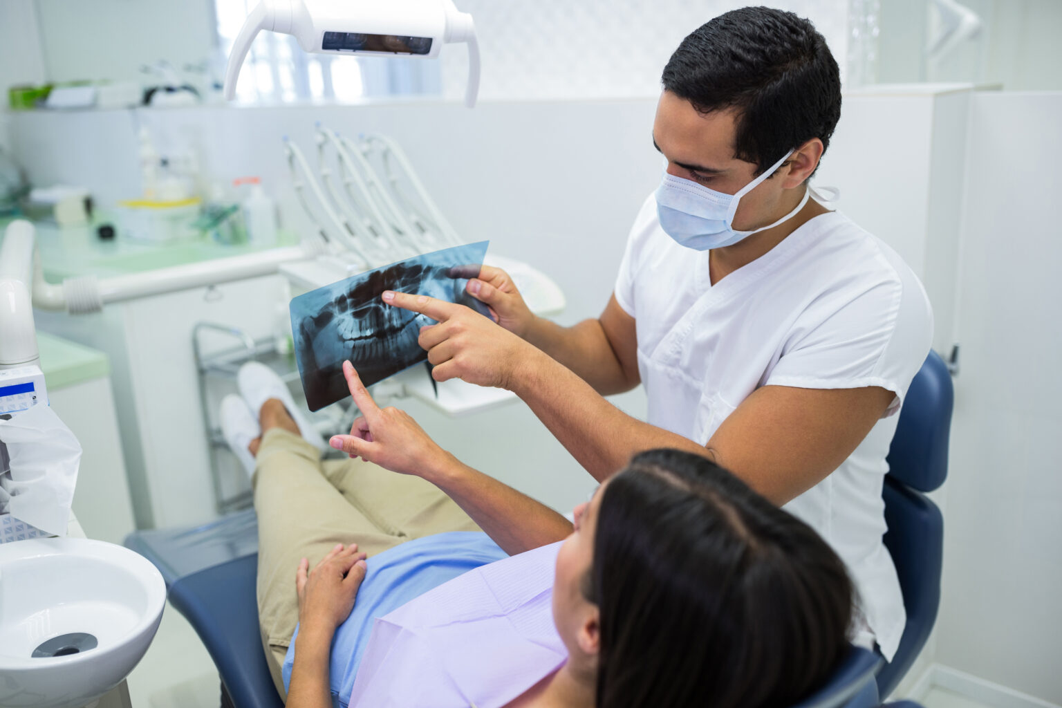
It is important to note that radiological examination, especially in the format of 3D tomography, plays a key role in determining the need and feasibility of sinus lifting, as well as in planning the operation itself.
Sinus lifting is an effective method that allows patients with insufficient bone volume in the upper jaw to receive implants and enjoy a beautiful smile and comfortable chewing function.
Operation Procedure
Lateral sinus lifting is performed under local anesthesia, which is a significant advantage for the patient since general anesthesia can negatively affect organs, especially the brain. This reduces operation time, simplifies the technique, and lowers the cost. The nerves of the upper jaw are blocked by locally injecting anesthetic near nerve nodes and trunks (conduction anesthesia).
Once anesthesia takes effect, a lateral approach to the maxillary sinus is made. The bone is drilled with a burr, or, less commonly, ultrasound or a scraper is used. Drilling provides good results, making the procedure less traumatic than scraping and more precise than ultrasound.
Next, a small window is created in the bone above the implant site, without affecting the sinus membrane. The operation should be performed by an experienced dental surgeon well-versed in anatomy. Quality instruments are used to avoid accidental damage.
After the operation, X-rays show the sinus membrane behind the removed bone. The gap is filled with osteoplastic material that stimulates new bone formation. A good option is the patient’s ground bone tissue, but biological or synthetic materials are often required.
The window in the upper jaw is closed, sometimes using barrier membranes or plates. The wound is sutured, and the postoperative period usually passes without serious complications. The patient is given recommendations regarding diet and care.
Complications
Complications can include sinus membrane perforation, operation inefficiency, and sinusitis exacerbation. Small windows are prone to perforation, and inefficiency may be caused by unsuccessful bone tissue regeneration. Sinusitis is treated with standard methods.
Conclusion
Despite the challenges and potential complications, sinus lifting is a safe and effective operation that provides patients with a white smile and the ability to comfortably eat.
Часто задаваемые вопросы
Какова опасность этого заболевания?
Заболевание вызывается кариозными бактериями, которые вырабатывают кислоты, постепенно разрушая твердые ткани зубов и делая их хрупкими из-за потери кальция и фтора. Если этот процесс не остановить своевременно, зуб обязательно разрушится.
Почему появляется кариес на зубах?
Основной фактор – неправильная и нерегулярная чистка зубов. Эмаль, а затем дентин (твердая ткань зуба) разрушаются под воздействием кислот, выделяемых бактериями в ротовой полости.
Как осуществляется удаление кариеса на стадии пятна?
Если поражение незначительное, удаление кариозных областей может и не потребоваться. Во многих случаях антисептическая (медикаментозная) обработка и применение реминерализирующего геля с кальцием и фтором достаточны для восстановления эмали.
Как можно избавиться от кариеса дома?
В настоящее время нет средств для домашнего лечения. Для эффективного лечения необходимы специальное оборудование, антисептические препараты и материалы для восстановления твердых тканей.
Сколько времени занимает лечение?
Время лечения зависит от степени поражения. На поверхностной стадии обычно требуется 10-25 минут, на средней – 30-60 минут, при значительных повреждениях процедуры могут занять до полутора часов.
Болезненно ли лечение?
Лечение начального кариеса обычно безболезненно. На более поздних стадиях применяется анестезия. Десна предварительно обрабатывают аппликационным анестетиком, поэтому даже укол не вызывает дискомфорта.
В каких случаях лечение противопоказано?
Процедура почти не имеет противопоказаний, за исключением пациентов с тяжелыми заболеваниями центральной нервной системы. При обострении сердечно-сосудистых патологий и тяжелых заболеваниях дыхательных путей лечение лучше перенести на более поздний срок.
Можно ли лечить кариес во время беременности?
Беременным разрешено лечить только поверхностный кариес без ограничений. Использование анестезии допускается только во втором триместре. При возможности рекомендуется отложить процедуру до рождения ребенка.
Что включает в себя профилактика этого заболевания?
Эффективная домашняя гигиена, включая чистку зубов дважды в день и использование ополаскивателей после еды. Ограничение потребления сладкой и углеводной пищи. Регулярное посещение стоматолога для профилактической гигиенической обработки ротовой полости каждые 4-6 месяцев.
